Definition
Focal atrial tachycardia (FAT) is a form of supraventricular tachycardia (SVT) originating from a single ectopic focus within the atria but outside of the sinus node
- The term FAT is commonly used synonymously with atrial tachycardia, a broader term referring to any form of SVT originating within the atria but outside of the sinus node
- FAT, atrial flutter and multifocal atrial tachycardia (MAT) are all forms of atrial tachycardia
- Management of the three types varies and thus distinguishing between them is clinically important
Pathophysiology of FAT
- Due to a single ectopic focus
- The underlying mechanism can involve increased automaticity, triggered activity or reentry
- May be paroxysmal or sustained
- Multiple causes including:
- Digoxin toxicity
- Atrial scarring due to ischaemic heart disease
- Catecholamine excess
- Stimulants including cocaine, caffeine
- Alcohol
- Congenital abnormalities
- Idiopathic
- Sustained atrial tachycardia may rarely be seen and can progress to tachycardia-induced cardiomyopathy
ECG Features of Atrial Tachycardia
- Atrial rate > 100 bpm
- Abnormal P wave morphology and axis (e.g. inverted in inferior leads) due to ectopic origin
- Unifocal, identical P waves
- Isoelectric baseline (unlike atrial flutter)
- Normal QRS morphology (unless pre-existing bundle branch block, accessory pathway, or rate-related aberrant conduction)
AV block may be present — this is generally a physiological response to the rapid atrial rate, except in digoxin toxicity where there is AV nodal suppression due to vagotonic effects of digoxin, resulting in a slow ventricular rate (“PAT with block”).
ECG Examples
Example 1
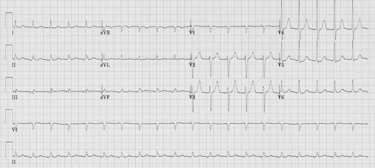
Focal atrial tachycardia:
- There is a narrow complex tachycardia at 120 bpm
- Each QRS complex is preceded by an abnormal P wave — upright in V1, inverted in the inferior leads II, III and aVF
- P wave morphology is consistent throughout
Example 2
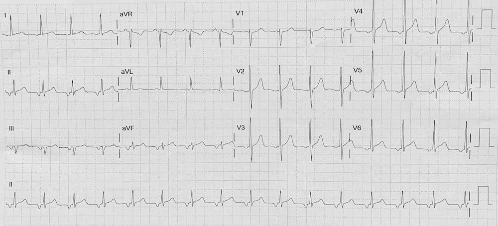
Focal atrial tachycardia:
- There is a narrow complex tachycardia at 95 bpm
- Each QRS complex is preceded by an abnormal P wave — biphasic in V1; inverted in the inferior leads II, III and aVF; and inverted V3-V6
- P wave morphology is consistent throughout
Related Topics
References
Advanced Reading
Online
Textbooks
- Zimmerman FH. ECG Core Curriculum. 2023
- Mattu A, Berberian J, Brady WJ. Emergency ECGs: Case-Based Review and Interpretations, 2022
- Straus DG, Schocken DD. Marriott’s Practical Electrocardiography 13e, 2021
- Brady WJ, Lipinski MJ et al. Electrocardiogram in Clinical Medicine. 1e, 2020
- Mattu A, Tabas JA, Brady WJ. Electrocardiography in Emergency, Acute, and Critical Care. 2e, 2019
- Hampton J, Adlam D. The ECG Made Practical 7e, 2019
- Kühn P, Lang C, Wiesbauer F. ECG Mastery: The Simplest Way to Learn the ECG. 2015
- Grauer K. ECG Pocket Brain (Expanded) 6e, 2014
- Surawicz B, Knilans T. Chou’s Electrocardiography in Clinical Practice: Adult and Pediatric 6e, 2008
- Chan TC. ECG in Emergency Medicine and Acute Care 1e, 2004
LITFL Further Reading
Emergency Physician in Prehospital and Retrieval Medicine in Sydney, Australia. He has a passion for ECG interpretation and medical education | ECG Library |
MBBS DDU (Emergency) CCPU. Adult/Paediatric Emergency Medicine Advanced Trainee in Melbourne, Australia. Special interests in diagnostic and procedural ultrasound, medical education, and ECG interpretation. Co-creator of the LITFL ECG Library. Twitter: @rob_buttner