- Narrow Complex Tachycardia
- Ventricular rate 150bpm
Other differentials include AVNRT / AVRT however the rate is usually higher in these.
‘Mapping’ of flutter waves may be helpful, this may be easier if paper speed is altered e.g. 50mm/sec
Trial of vagal maneuvers of adenosine may help differentiate Atrial Flutter
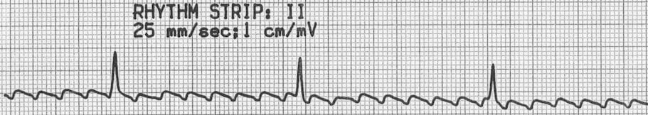
This patient received adenosine, rhythm strip below, revealing obvious flutter waves. In comparison to ECG Quiz 017, this patient does not cardiovert to sinus rhythm following an adenosine bolus. Instead, the degree of AV block is transiently increased, revealed underlying flutter waves and confirming the diagnosis of atrial flutter with a 2:1 block.
Would the Lewis Lead have helped?
The Lewis lead configuration can help to detect atrial activity and its relationship to ventricular activity. Useful in:
- Observing flutter waves in atrial flutter
- Detecting P waves in wide complex tachyarrhythmia to identify atrioventricular dissociation
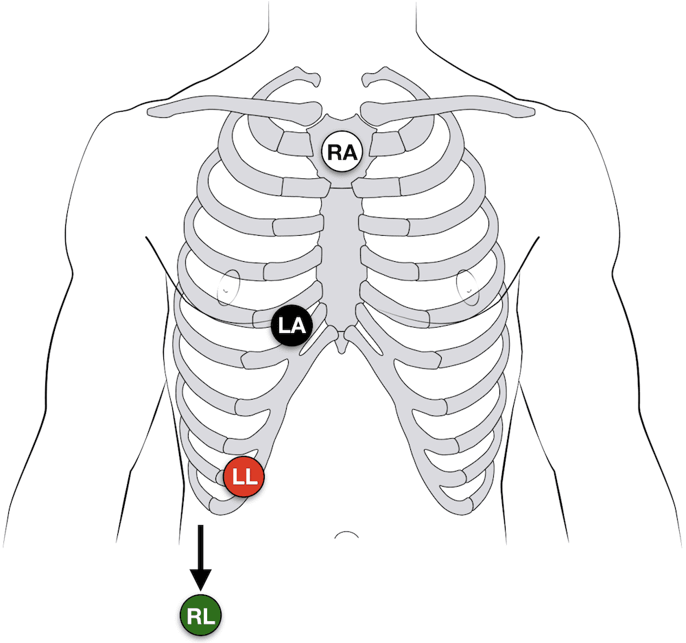
Lewis lead placement
- Right Arm (RA)electrode on manubrium
- Left Arm (LA) electrode over 5th ICS, right sternal border.
- Left Leg (LL) electrode over right lower costal margin.
- Monitor Lead I