30-year old Thai male presenting with syncope. Describe the ECG.
Describe and interpret this ECG
ECG ANSWER and INTERPRETATION
ECG Findings
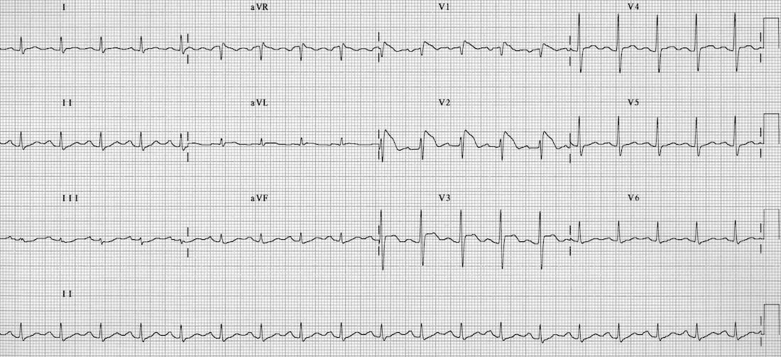
The patient is in sinus rhythm with no evidence of dysrhythmia or AV block.
The QT interval is normal and there is no evidence of WPW syndrome, HOCM or ARVD.
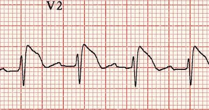
There is a characteristic pattern of abnormalities in V1-2:
- RSR’ pattern / partial RBBB
- ST elevation with a “coved” morphology
- Inversion of the terminal portion of the T wave
In a patient presenting with syncope, this ECG is diagnostic of the Brugada syndrome.
CLINICAL PEARLS
Syncope ECG Checklist
When faced with a patient presenting with syncope, systematically assess the ECG for the following abnormalities (click each item for details):
Emergency Physician in Prehospital and Retrieval Medicine in Sydney, Australia. He has a passion for ECG interpretation and medical education | ECG Library |
MBBS DDU (Emergency) CCPU. Adult/Paediatric Emergency Medicine Advanced Trainee in Melbourne, Australia. Special interests in diagnostic and procedural ultrasound, medical education, and ECG interpretation. Co-creator of the LITFL ECG Library. Twitter: @rob_buttner