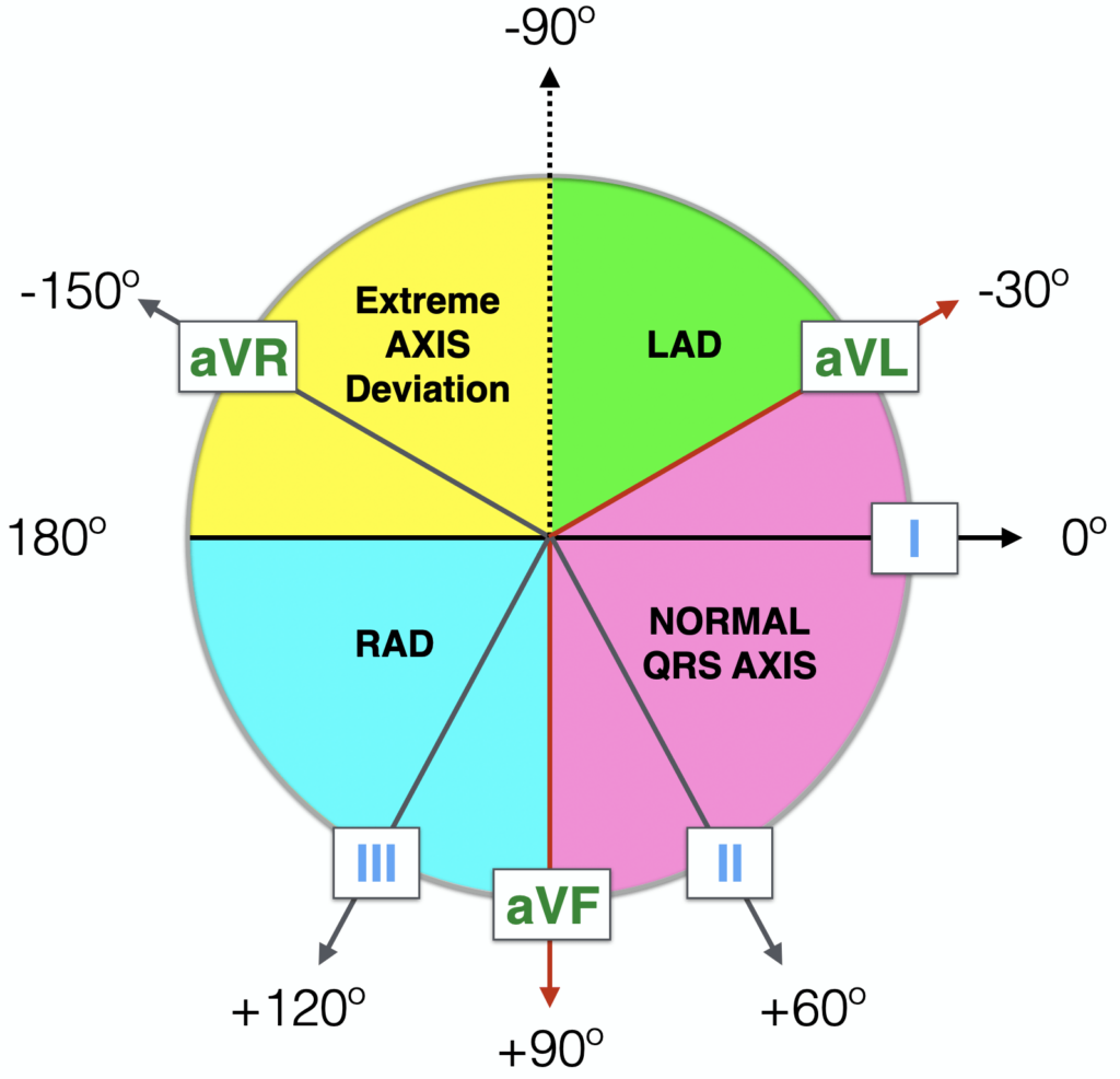
Left Axis Deviation LAD
Left Axis Deviation = QRS axis less than -30°.
- Normal Axis = QRS axis between -30° and +90°
- Right Axis Deviation = QRS axis greater than +90°
- Extreme Axis Deviation = QRS axis between -90° and 180° (AKA “Northwest Axis”)
Hexaxial Reference System
How to recognise left axis deviation
Three Lead analysis
- QRS is POSITIVE (dominant R wave) in Lead I
- QRS is NEGATIVE (dominant S wave) in leads II, III and aVF
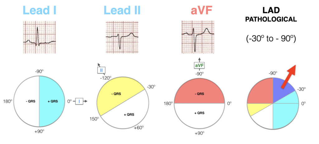
Example ECG of LAD

Left Axis Deviation:
- Leads I and aVL are positive; leads II and aVF are negative
Causes of LAD
Advanced Reading
Online
Textbooks
- Zimmerman FH. ECG Core Curriculum. 2023
- Mattu A, Berberian J, Brady WJ. Emergency ECGs: Case-Based Review and Interpretations, 2022
- Straus DG, Schocken DD. Marriott’s Practical Electrocardiography 13e, 2021
- Brady WJ, Lipinski MJ et al. Electrocardiogram in Clinical Medicine. 1e, 2020
- Mattu A, Tabas JA, Brady WJ. Electrocardiography in Emergency, Acute, and Critical Care. 2e, 2019
- Hampton J, Adlam D. The ECG Made Practical 7e, 2019
- Kühn P, Lang C, Wiesbauer F. ECG Mastery: The Simplest Way to Learn the ECG. 2015
- Grauer K. ECG Pocket Brain (Expanded) 6e, 2014
- Surawicz B, Knilans T. Chou’s Electrocardiography in Clinical Practice: Adult and Pediatric 6e, 2008
- Chan TC. ECG in Emergency Medicine and Acute Care 1e, 2004
LITFL Further Reading
Emergency Physician in Prehospital and Retrieval Medicine in Sydney, Australia. He has a passion for ECG interpretation and medical education | ECG Library |
MBBS DDU (Emergency) CCPU. Adult/Paediatric Emergency Medicine Advanced Trainee in Melbourne, Australia. Special interests in diagnostic and procedural ultrasound, medical education, and ECG interpretation. Co-creator of the LITFL ECG Library. Twitter: @rob_buttner