Elderly patient presenting with nausea and visual disturbance. Interpret the ECG.
Describe and interpret this ECG
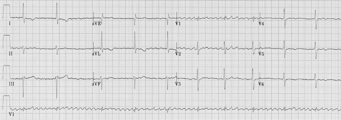
ECG ANSWER and INTERPRETATION
This is a tricky ECG!
There is evidence of atrial fibrillation, as evidenced by the irregular baseline with fibrillatory waves most prominent in V1-2.
NB. Fibrillatory waves are characteristically seen in V1-2 (which overlie the atria), as opposed to tremor artefact which may be in seen in multiple leads without a predominance for V1-2.
However, the ventricular rhythm is regular. How can this be? AF is irregular by definition…
This is an example of “regularised AF” due to digoxin toxicity:
If this all seems like too much of a coincidence, then consider the pathophysiology of digoxin toxicity…
CLINICAL PEARLS
Mechanisms of Digoxin Toxicity
Digoxin toxicity produces a wide variety of dysrhythmias, due to:
- Increased automaticity of atrial, junctional and
ventricular tissues — via actions at the Na/K and Na/Ca exchangers
causing increased intracellular calcium and therefore increased
spontaneous depolarisation of cardiac pacemaker cells. - Decreased AV conduction — via increased vagal tone at the AV node.
Digoxin toxicity produces some combination of:
Characteristic ECG patterns include:
NB. Digoxin toxicity should not be confused with digoxin effect (= “sagging” ST depression and T-wave inversion in patients on therapeutic doses of digoxin; not predictive of toxicity).
Clinical Pearls
- Check for tremor artefact before you start diagnosing regularised AF!
- If the ECG pattern appears genuine and the clinical picture is
compatible with digoxin toxicity (GI upset, xanthopsia, current digoxin
treatment), then check an urgent digoxin level.
Emergency Physician in Prehospital and Retrieval Medicine in Sydney, Australia. He has a passion for ECG interpretation and medical education | ECG Library |