Poor R Wave Progression Overview
Poor R-wave progression (PRWP) is a common ECG finding that is often inconclusively interpreted as suggestive, but not diagnostic, of anterior myocardial infarction (AMI). PRWP is defined by R wave height ≤ 3 mm in V3
Causes of PRWP
The major causes associated with PRWP include:
ECG Examples
Example 1
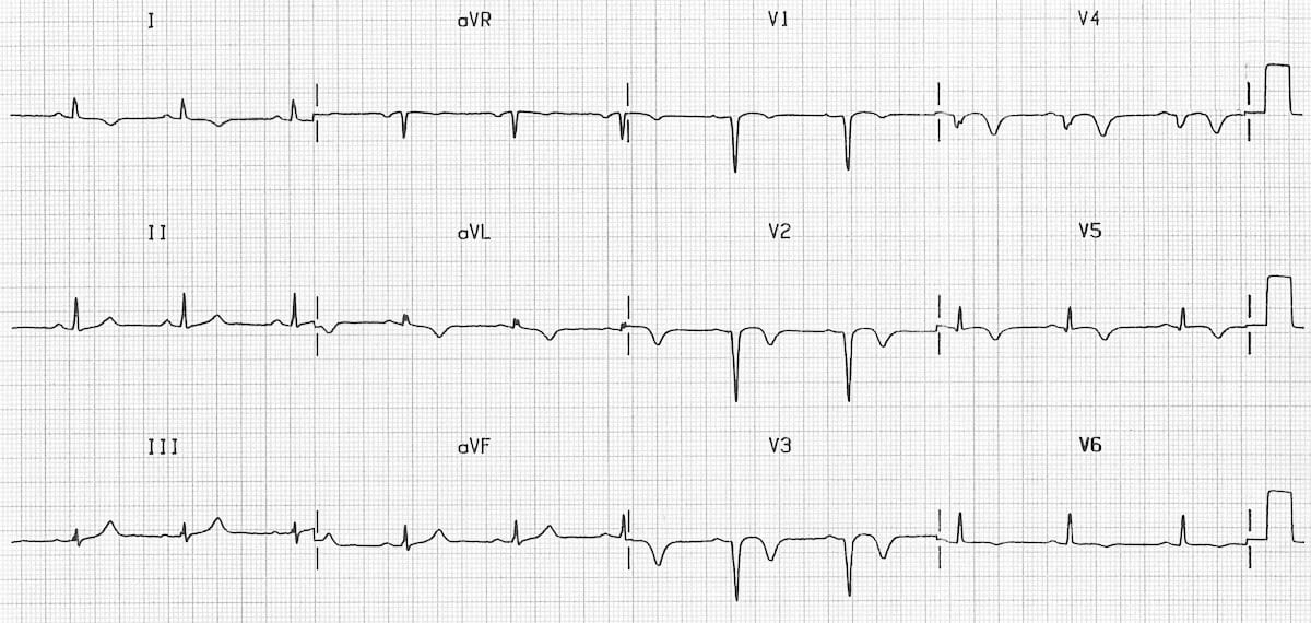
Poor R wave progression secondary to prior anteroseptal MI
Example 2
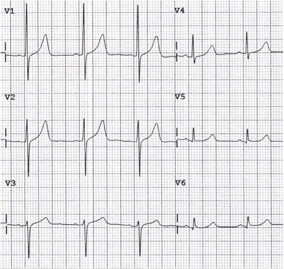
Poor R wave progression due to electrode misplacement (leads V1 and V3 reversed)
References
- Zema MJ, Kligfield P. ECG poor R-wave progression: review and synthesis. Arch Intern Med. 1982 Jun;142(6):1145-8. [PMID 6212033]
Advanced Reading
Online
Textbooks
- Zimmerman FH. ECG Core Curriculum. 2023
- Mattu A, Berberian J, Brady WJ. Emergency ECGs: Case-Based Review and Interpretations, 2022
- Straus DG, Schocken DD. Marriott’s Practical Electrocardiography 13e, 2021
- Brady WJ, Lipinski MJ et al. Electrocardiogram in Clinical Medicine. 1e, 2020
- Mattu A, Tabas JA, Brady WJ. Electrocardiography in Emergency, Acute, and Critical Care. 2e, 2019
- Hampton J, Adlam D. The ECG Made Practical 7e, 2019
- Kühn P, Lang C, Wiesbauer F. ECG Mastery: The Simplest Way to Learn the ECG. 2015
- Grauer K. ECG Pocket Brain (Expanded) 6e, 2014
- Surawicz B, Knilans T. Chou’s Electrocardiography in Clinical Practice: Adult and Pediatric 6e, 2008
- Chan TC. ECG in Emergency Medicine and Acute Care 1e, 2004
LITFL Further Reading
Emergency Physician in Prehospital and Retrieval Medicine in Sydney, Australia. He has a passion for ECG interpretation and medical education | ECG Library |
MBBS DDU (Emergency) CCPU. Adult/Paediatric Emergency Medicine Advanced Trainee in Melbourne, Australia. Special interests in diagnostic and procedural ultrasound, medical education, and ECG interpretation. Co-creator of the LITFL ECG Library. Twitter: @rob_buttner